Videos


Images
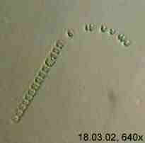
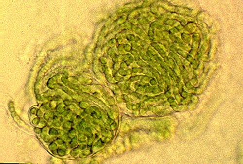
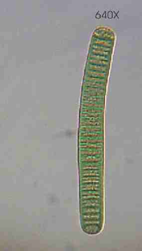
Anabaena affinis, 640X
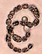
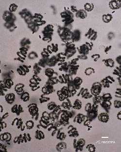
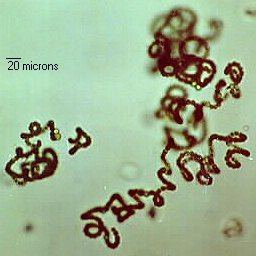
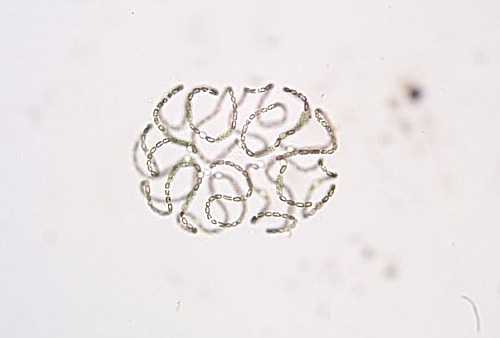
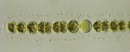
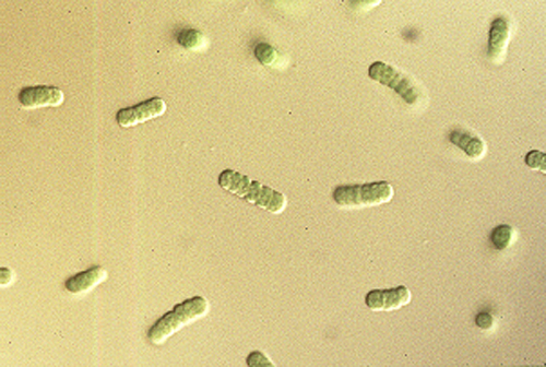
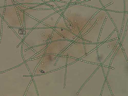
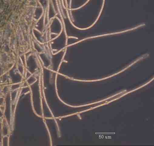
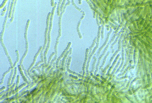
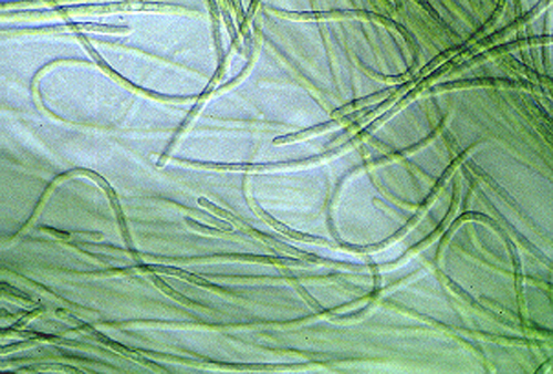
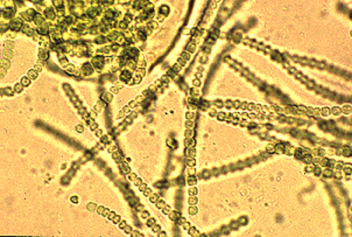
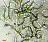
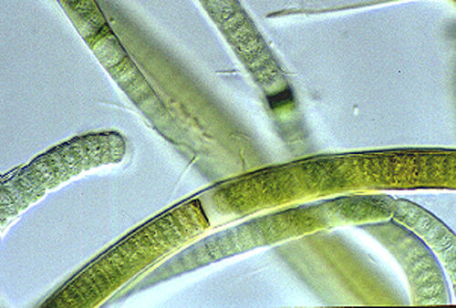
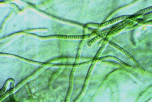
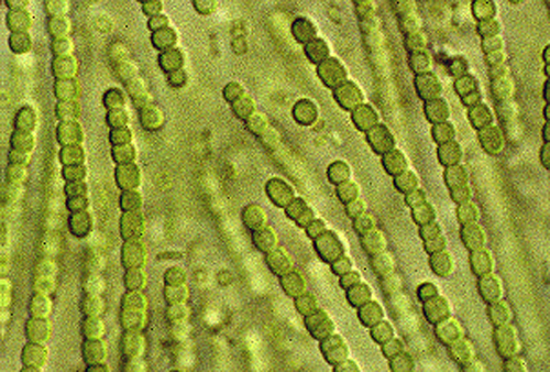
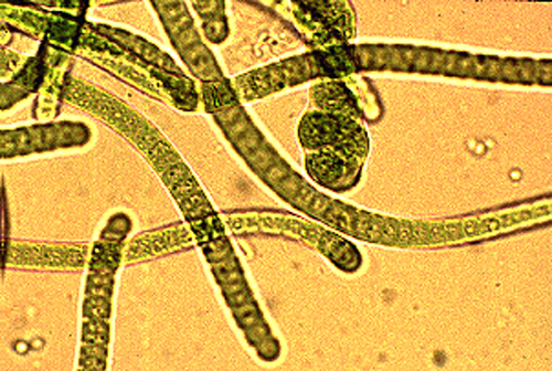
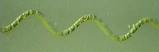
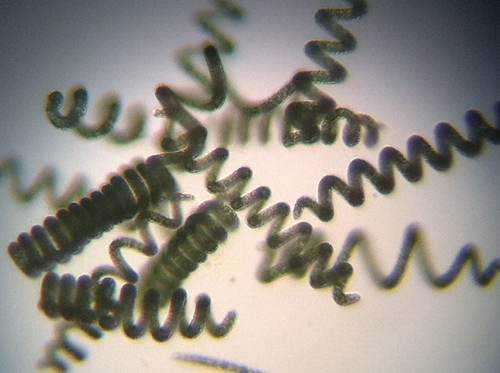
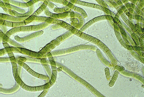
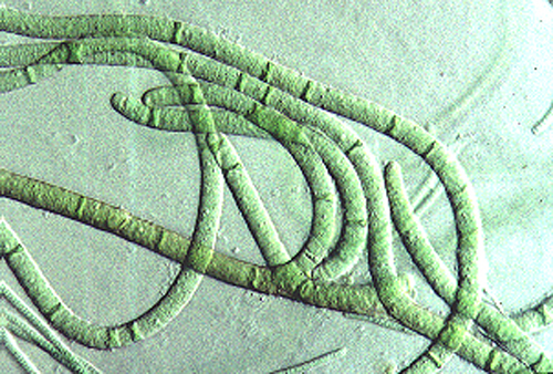
Anabaena circinalis - Measurement unavailable. probably 400X

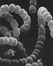
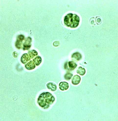
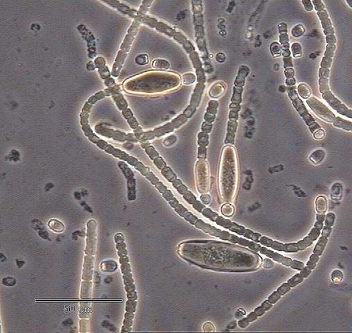
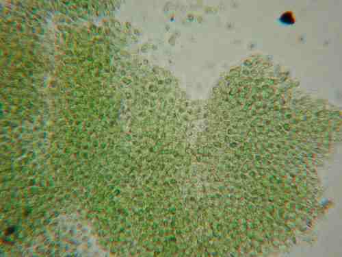
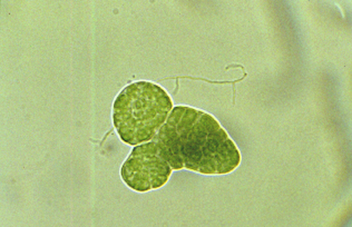
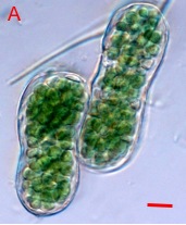
Anabaena flos-aquae enlarged 2500X

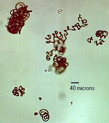
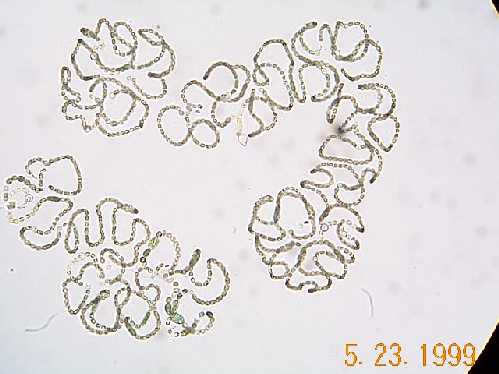
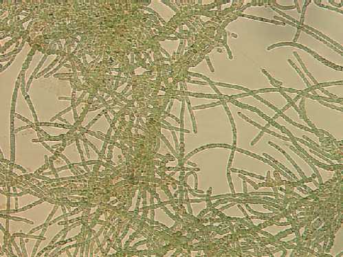
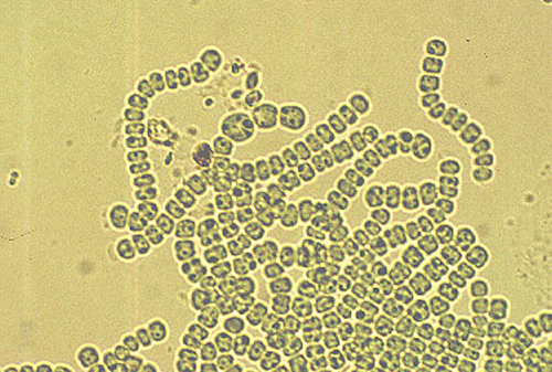
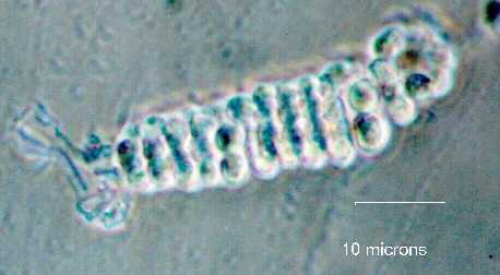
Anabaena "laxa" - ID for this complex is of course tenuous--see next entry. Measurement unavailable. probably 400X

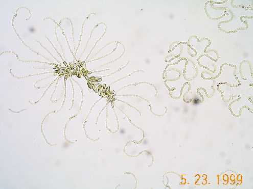
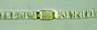
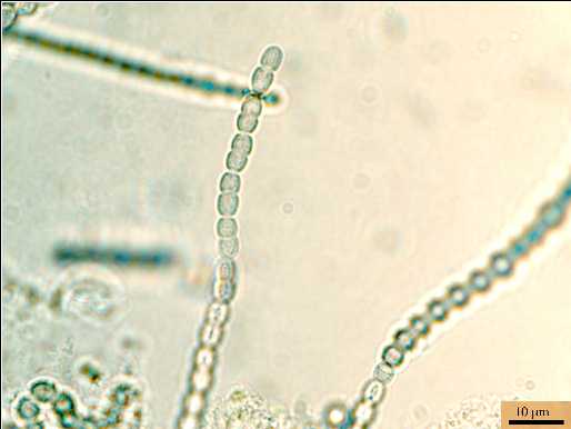
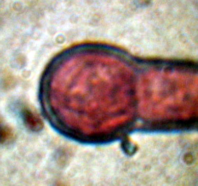
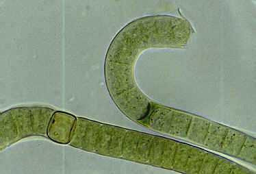
Anabaena "laxa" - From the same sample as above, a few weeks later. Note that the akinete is now adjacent the heterocyst. It now keys out to Anabaena "cylindrica". 400X
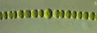
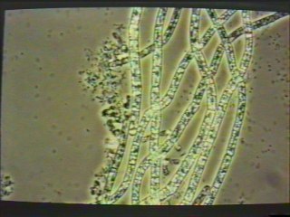
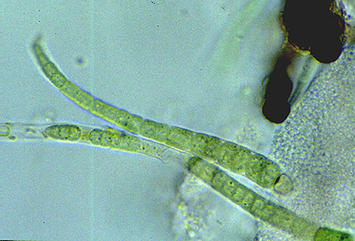
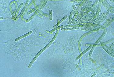
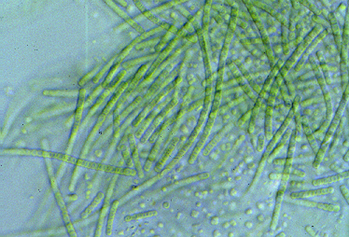
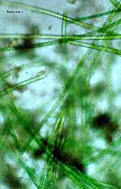
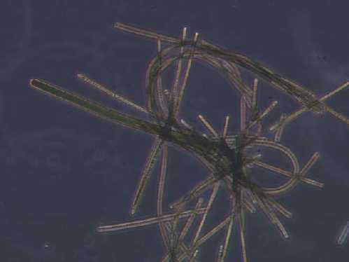
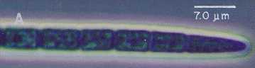
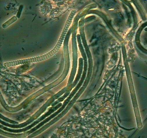
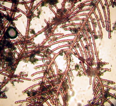
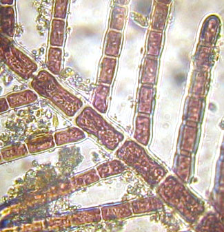
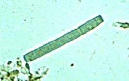
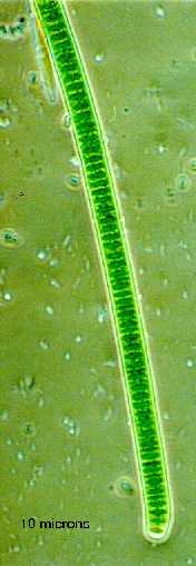
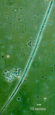
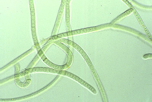
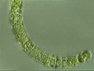
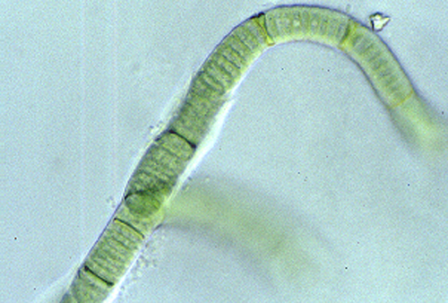
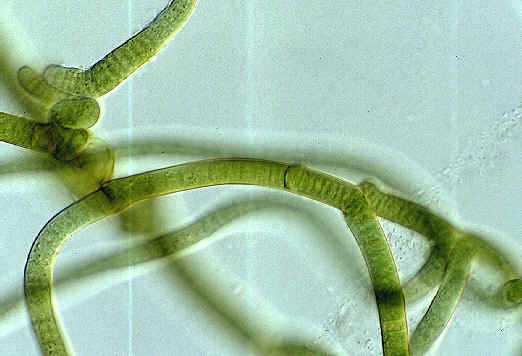
Anabaena scheremetievi - 400X phase contrast
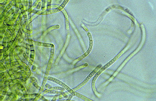
Anabaena scheremetievi - A slight hint of a sheath is visible. 400X

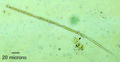
Anabaena scheremetievi - 400X
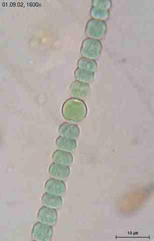
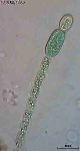
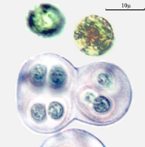
Anabaena sphaerica, 1600X
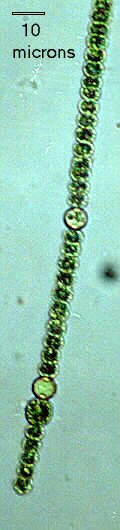
Anabaena sphaerica - 400X
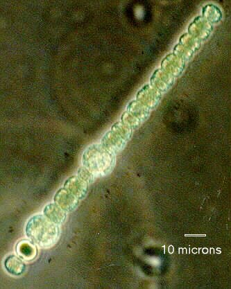
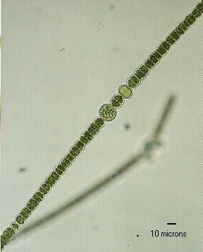
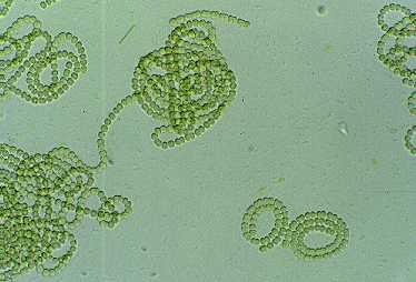
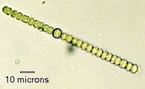
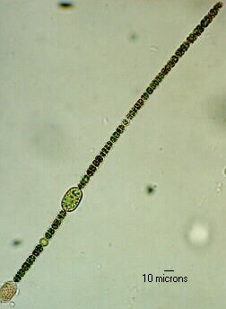
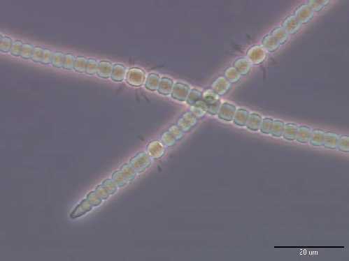
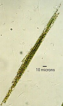
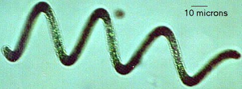
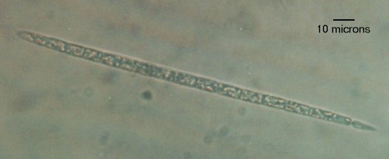
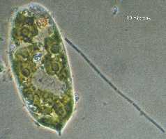
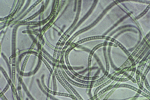
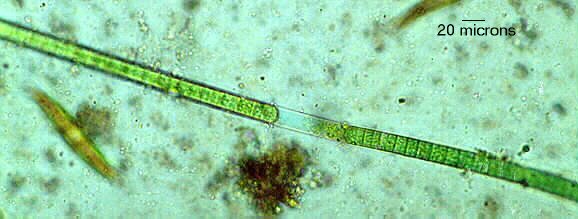
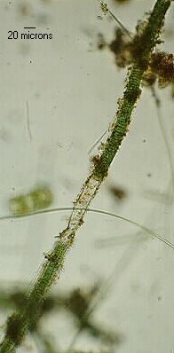
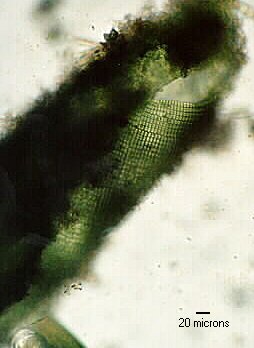
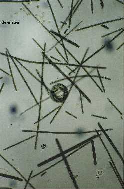
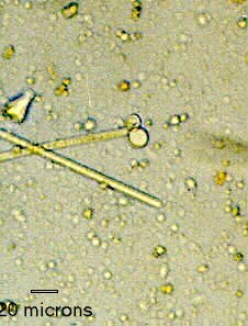
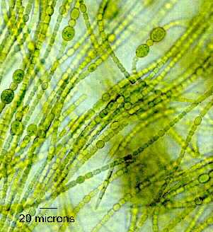
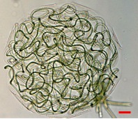
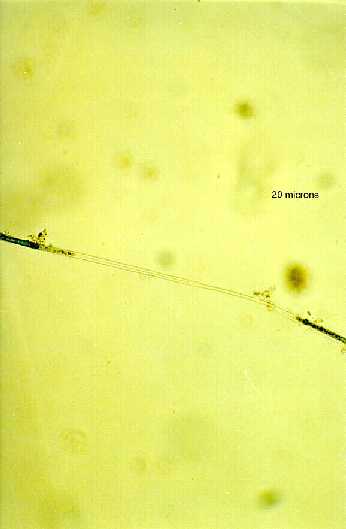
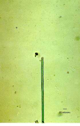
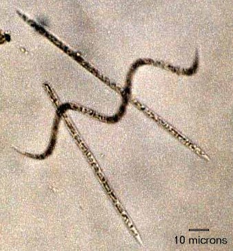
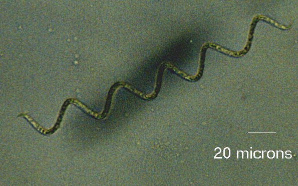
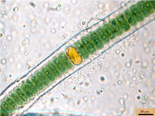
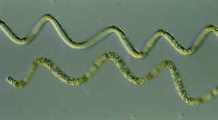
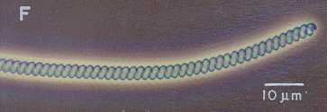
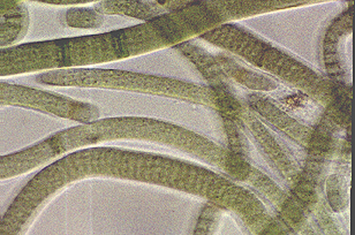
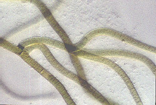
Anabaena spiroides - The line is 20 microns. 200X

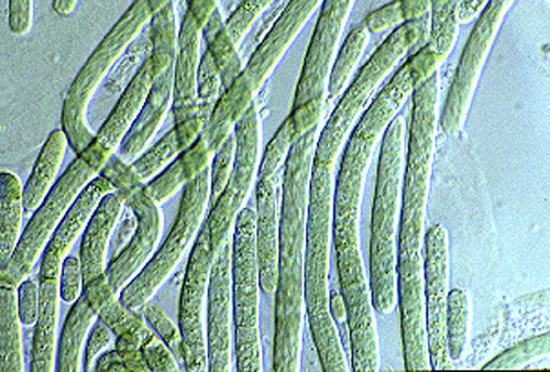
Anabaena torulosa - 400X

Anabaena torulosa - Is it Synecococcus? No, it is Anabaena torulosa akinetes. 400X
Anabaena sp. - (Unidentified) No speculations upon the species. 400X
Anabaena sp. - (Unidentified) No speculations upon the species. 400X
Anabaena sp. - (Unidentified) No speculations upon the species. 400X
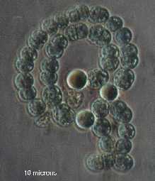
Anabaena sp. - (Unusual and contorted) Akinetes are round, so it is probably not A. circinalis. Could be A. spiroides in an unusual state. 200X.
Anabaena sp. - (Unusual and contorted) The same specimen as above at 100X.

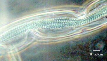
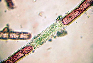
Anabaena sp. - Probably A. scheremetievi, but akinetes were absent. A hint of a sheath is barely visible. 400X
Anabaena sp. - Probably A. circinalis, but akinetes were absent. 1000X
Anabaena sp. - Possibly A. viguieri, but the akinetes are too broad. 400X.
Anabaena sp., from freshwater lake in Oregon, 100X
Anabaena sp., from freshwater lake in Oregon, 100X

Anabaena sp., from freshwater lake in Oregon, 200X
Anabaena sp., from freshwater lake in Oregon, 100X. Note the fabulous germinating akinetes.
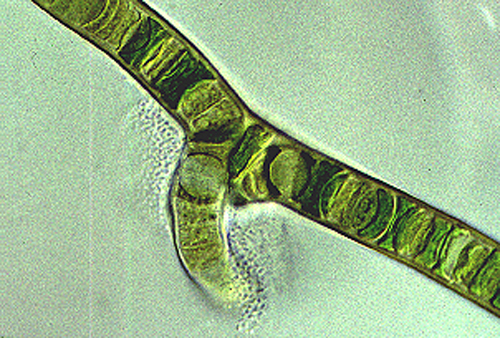
Anabaena sp. with symbiont bacteria (possibly Zoogloea) around heterocysts
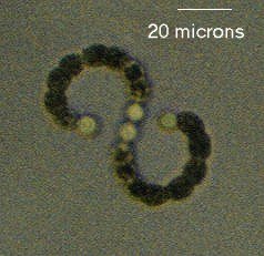
Anabaenopsis circularis - The central pair of heterocysts is clearly visible. 200X

Anabaenopsis circularis - A developing pair of heterocysts is visible in the center of the trichome. Note the bacteria attached to it. 400X (phase contrast)

Anabaenopsis circularis - Same as above without phase contrast. 400X
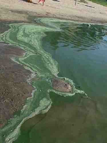
Anabaenopsis sp. bloom in Bedetti Lake, Santo Tome, Santa Fe, Argentina
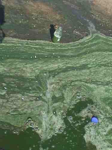
Anabaenopsis sp. bloom in Bedetti Lake, Santo Tome, Santa Fe, Argentina
Anabaenopsis sp. bloom in Bedetti Lake, Santo Tome, Santa Fe, Argentina
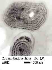
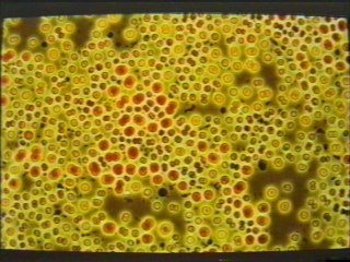
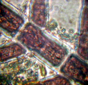
Anacystis nidulans TEM of semi-thin section (200 nm). See protocol.

Anacystis nidulans TEM of semi-thin section (200 nm) See protocol.
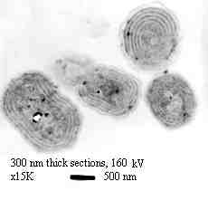
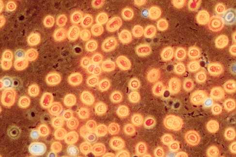
Anacystis nidulans TEM of semi-thin section (300 nm) See protocol.

Anacystis nidulans TEM of semi-thin section (300 nm) See protocol.

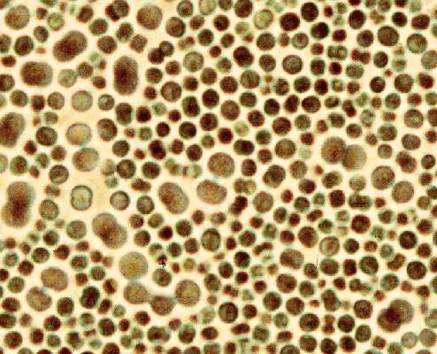
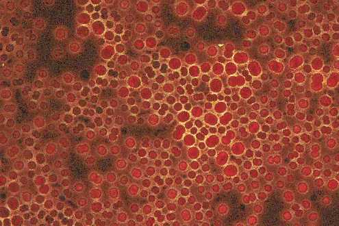
Anacystis nidulans TEM of semi-thin section (500 nm) See protocol.
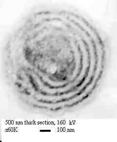
Anacystis nidulans TEM of semi-thin section (500 nm) See protocol.
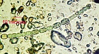
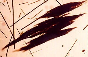
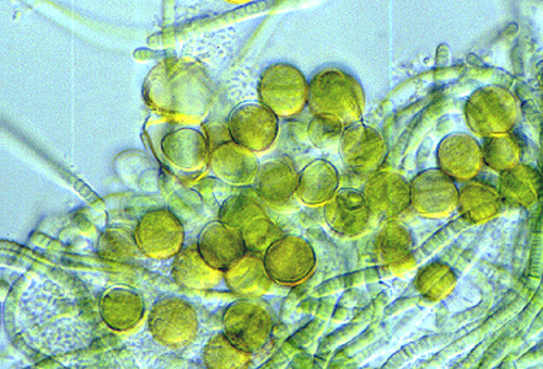
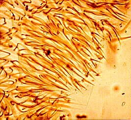
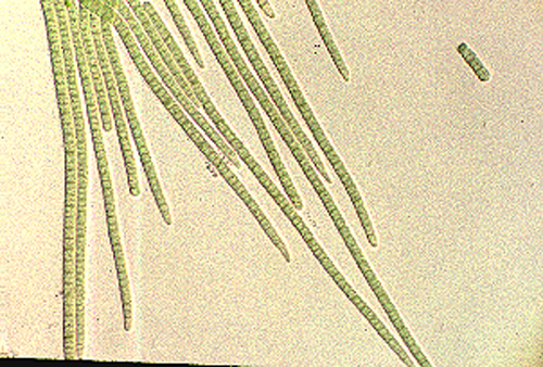
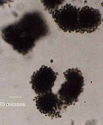
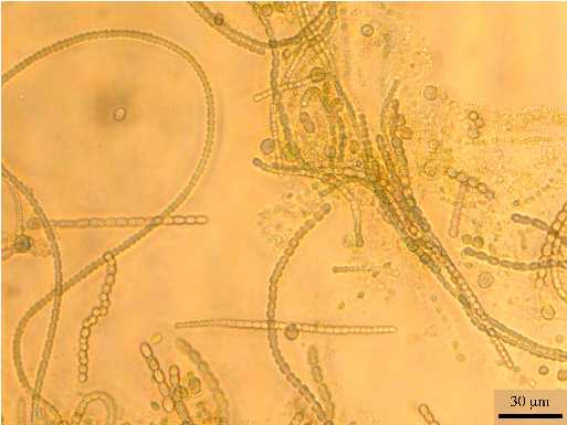
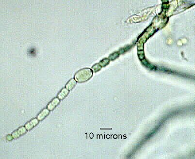
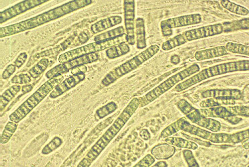
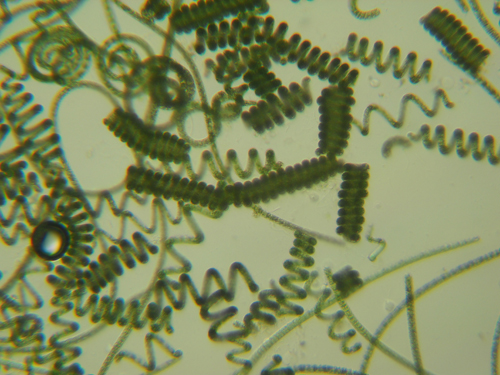
Aphanizomenon flos-aquae - Akinetes and heterocyst clearly visible. Measurement unavailable. probably 400X
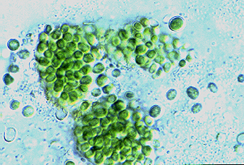
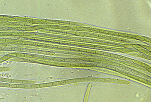
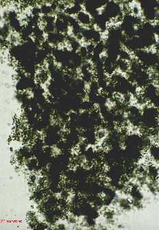
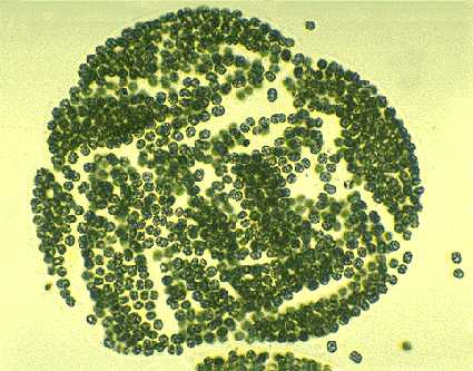
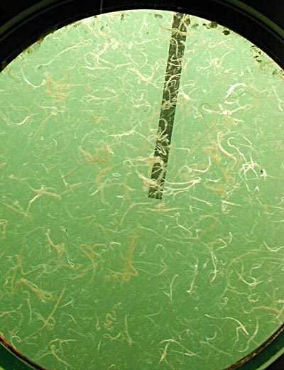
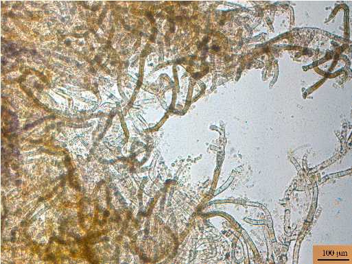
Aphanizomenon flos-aquae - Colony. Measurement unavailable. probably 100X
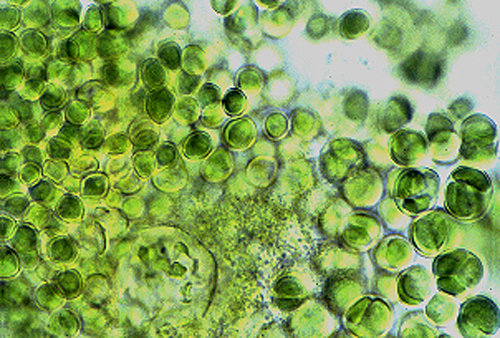
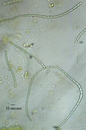
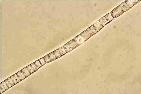
Aphanizomenon flos-aquae - Colony. Akinetes absent. 400X
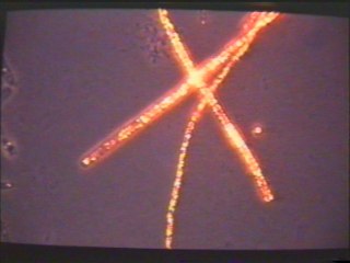
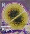
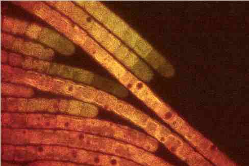
Aphanizomenon flos-aquae strain 1401/5. Autofluorescence image.

Aphanizomenon flos-aquae strain 1401/5. Autofluorescence image.
Aphanizomenon flos-aquae strain 1401/5. Autofluorescence image.

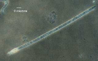
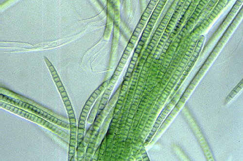
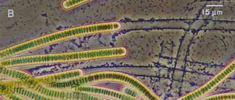
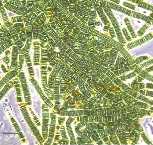
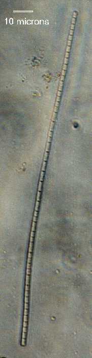
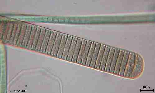
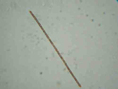
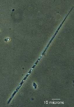
Arthrospira jenneri - Cross-walls obscured by cytoplasmic granules (sorry). 400X
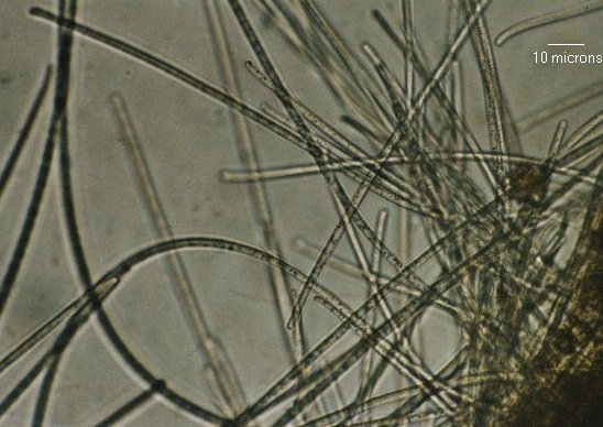
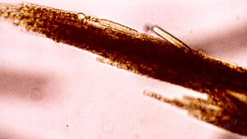
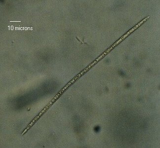
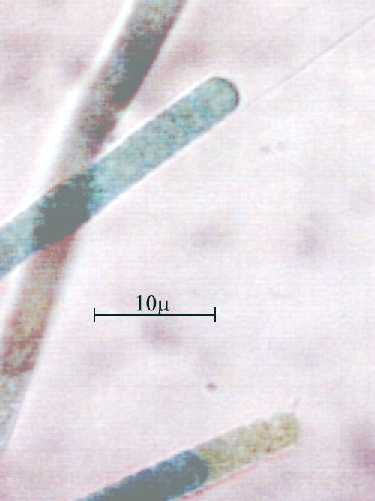
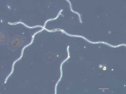
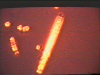
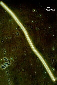
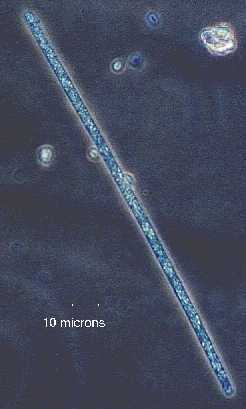
Beggiatoa alba - 400X
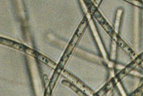
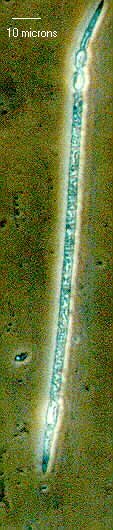
Beggiatoa alba - Cropped portion of image above at higher resolution.

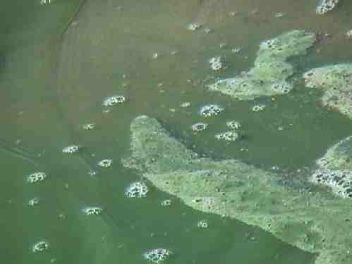
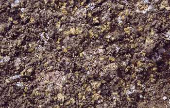
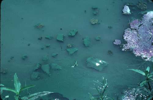
Bloom in waters at the Hartebeespoort dam in South Africa
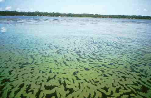
Bloom of Microcystis aeruginosa and Anabaena circinalis on the St. Johns River, FL

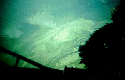
Bloom of Aphanizomenon flos aquae in a lake in Surrey, UK
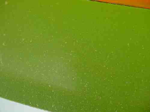

Bloom of Microcystis in Crescent Lake, FL

Bloom in Doctor's Lake
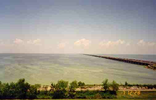
Bloom in Lake Ponchartrain, LA, an oligohaline estuary
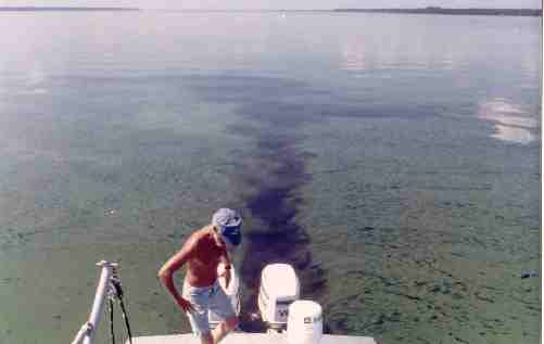
Bloom of Microcystis aeruginosa and Anabaena circinalis contaminated by a boat on the St. Johns River, FL
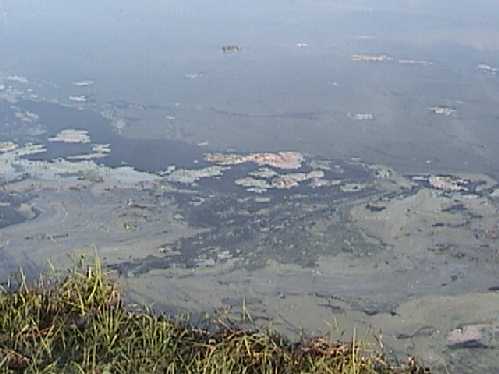
Bloom in agricultural setting in Missouri
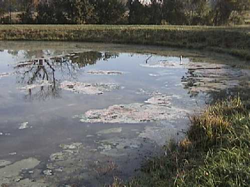
Bloom in agricultural setting in Missouri
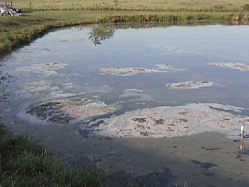
Bloom in agricultural setting in Missouri

Bloom in agricultural setting in Missouri
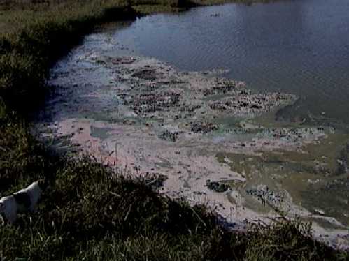
Bloom in agricultural setting in Missouri
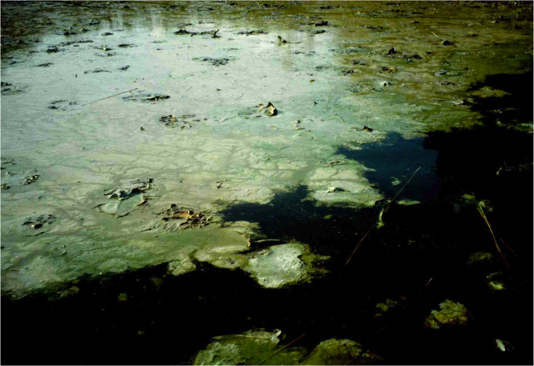
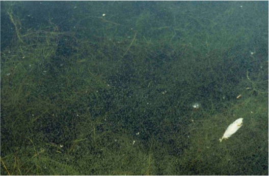
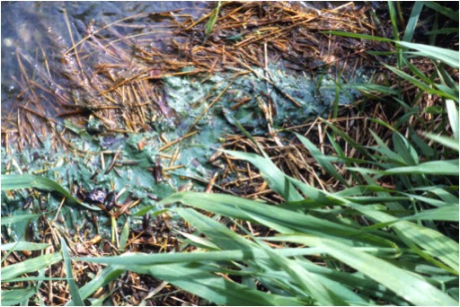
Anabaena and Aphanizomenon - decaying scum in a Scottish freshwater lake
Anabaena and Microcystis - bloom in a Scottish freshwater lake

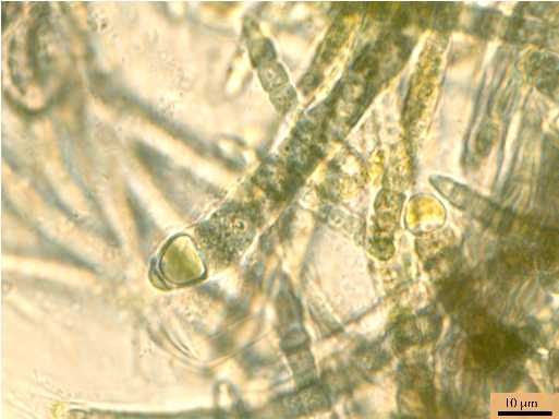

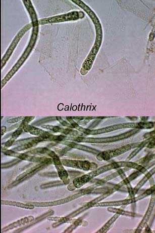
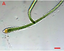
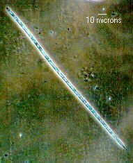
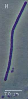
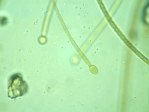
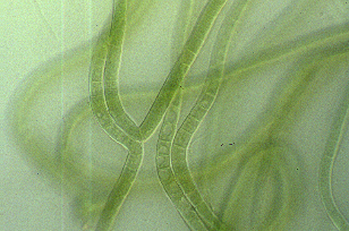
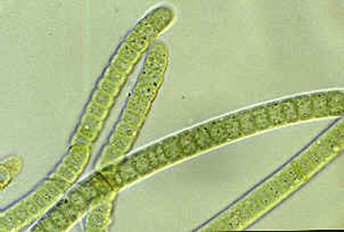
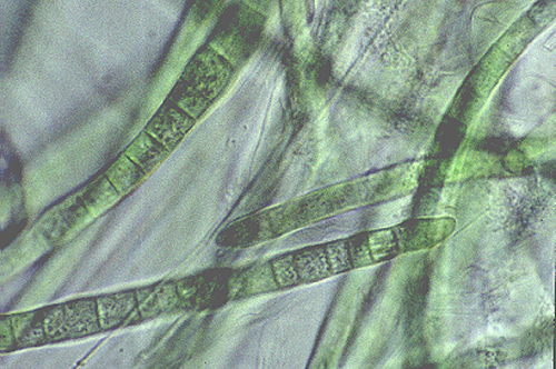
Calothrix sp. showing terminal heterocysts

Chloroflexus sp. - Unknown phytoplankton from a fish pond. Speculation: Chloroflexus. 400X
Chloroflexus sp. - Wider view higher resolution image of specimen above.
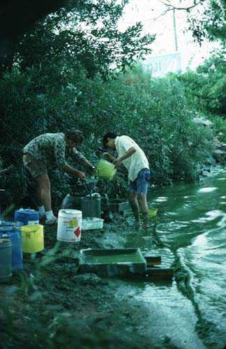
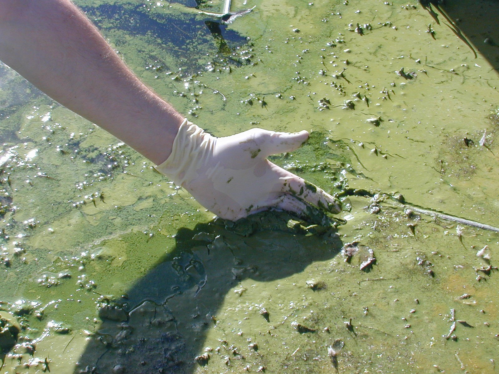
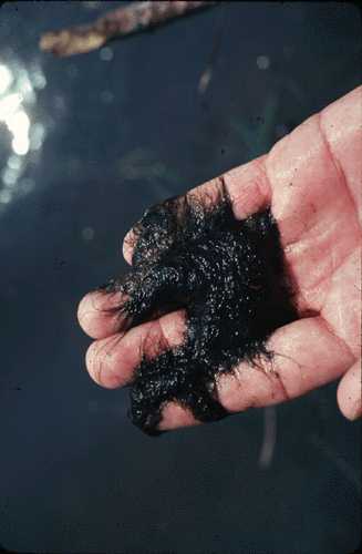
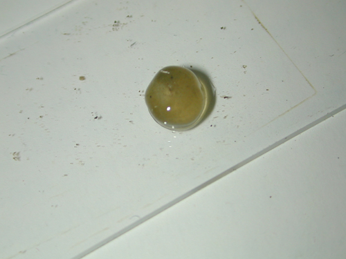
Collecting cyanobacteria from a large bloom
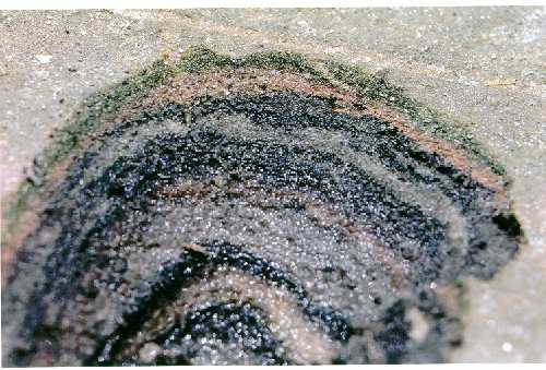
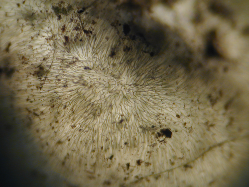
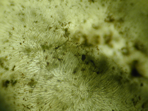
Cryptozoon proliferum - from Quebec

Cryptozoon proliferum - from Quebec

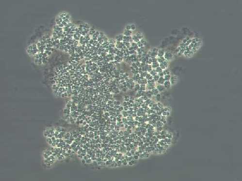
Cyanosarcina sp. from a pulp and paper waste-treatment system in Brazil
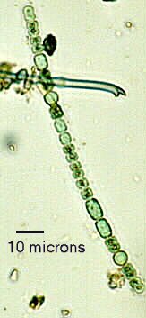
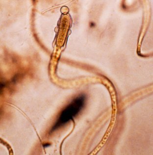
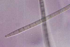
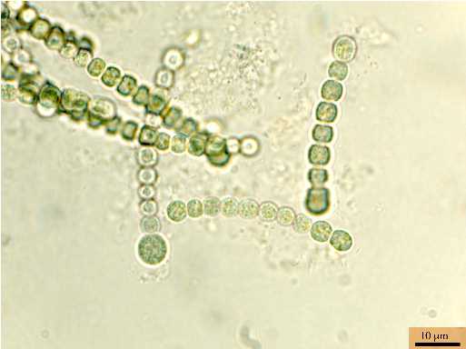
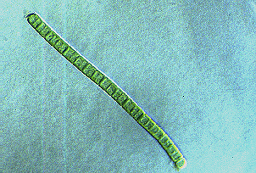
Cylindrospermopsis raciborskii - Heterocysts and akinetes visible. 400X
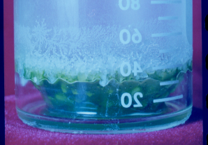
Very old laboratory culture of Cyanothece sp. forms solid roof to retain moisture.
Cyanothece sp. - electron micrograph showing carbohydrate granules
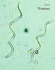
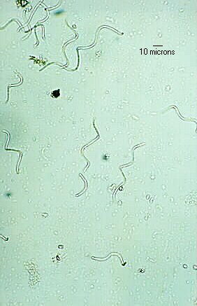
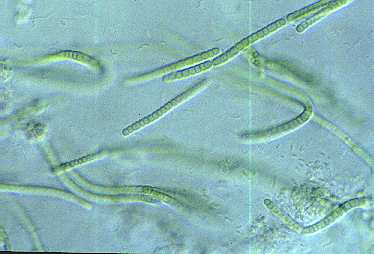
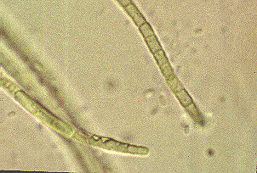
Cylindrospermopsis raciborskii - 400X phase contrast
Cylindrospermopsis raciborskii - Heterocyst visible. 400X
Cylindrospermopsis raciborskii - Akinetes (only) visible. 400X phase contrast
Cylindrospermopsis raciborskii - Heterocyst visible. 400X phase contrast
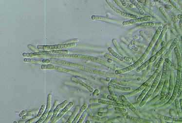
Cylindrospermum muscicola - 400X

Cylindrospermum muscicola - Surprise! It is not Pseudanabaena, but C. muscicola, without akinetes, but with a developing heterocyst. 400X phase contrast
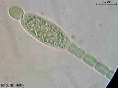
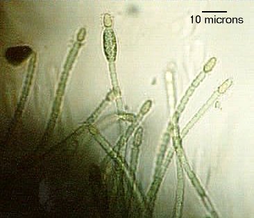
Cylindrospermum sp., 1600X
Cylindrospermum sp., 1600X
Eubacterium - Unidentified bacterium pays Euglena a visit. 1000X
Geitlerinema sp. from a pulp and paper waste-treatment system in Idaho
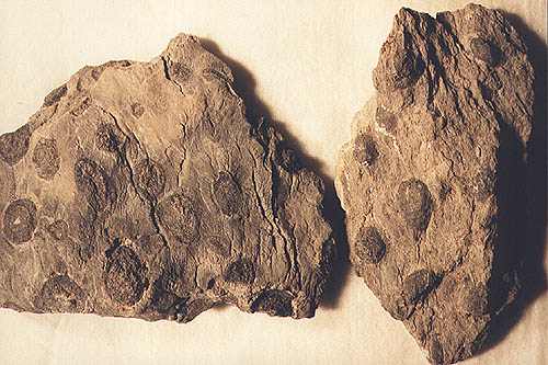
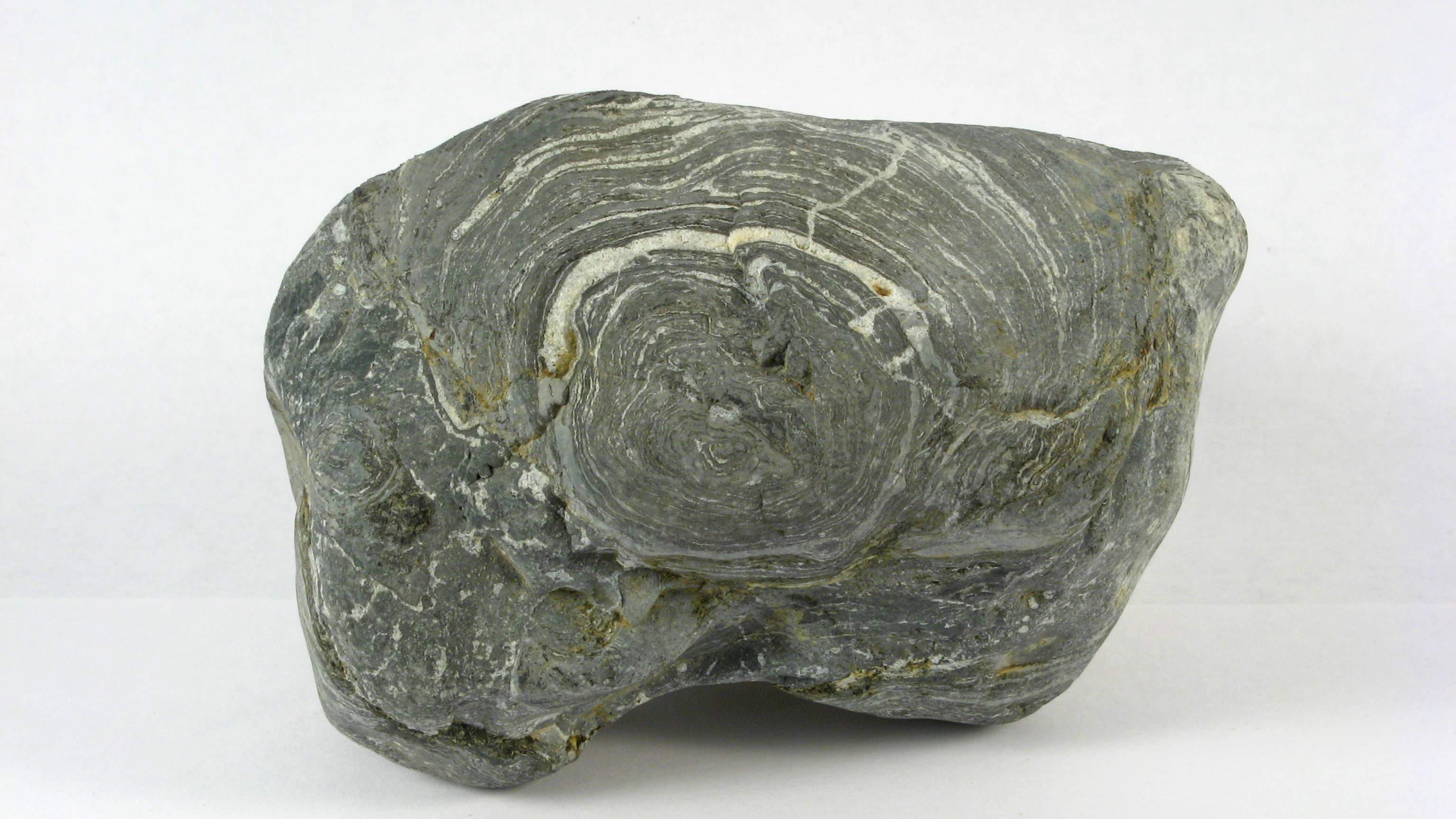
Girvanella sp. in algae oncolites from the Lower Cambrian Mule Spring Limestone, Waucoba Spring, Death Valley National Park
Gloeocapsa sp. together with two algae
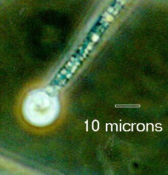
"Gloeotrichia" - Akinete and heterocyst clearly visible. Measurement unavailable. 400X
"Gloeotrichia" - Colony. Measurement unavailable. 100X
Gloeotrichia echinulata - pinhead-size colonies in a Scottish freshwater lake
Komvophoron sp.


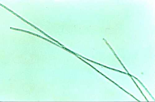
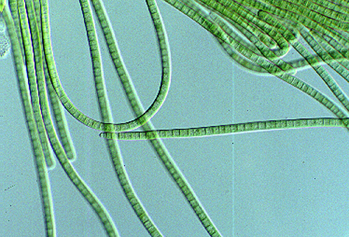
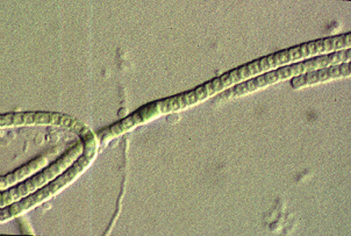
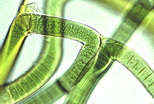
Lyngbya sp. showing classical sheath and terminal differentiation in the lower filament

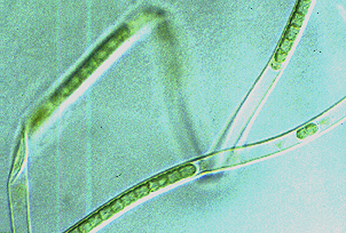
Lyngbya sp. showing formation of a coil at the end of a "hairpin" formation
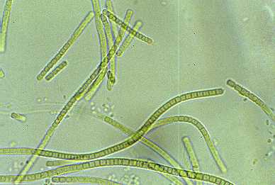
Lyngbya sp. - (Unidentified) No speculations upon the species. 200X

Lyngbya sp. - (Unidentified) No speculations upon the species. Note that one has no visible sheath. 200X
Lyngbya sp. - Same specimen as above. 400X phase contrast
Lyngbya sp. - (Unidentified) Possibly birgei. 200X
Lyngbya sp. - Social gathering of the above species. 200X

Lyngbya sp., two cells are leaving the shell

Lyngbya sp. at 100 X.
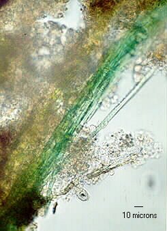
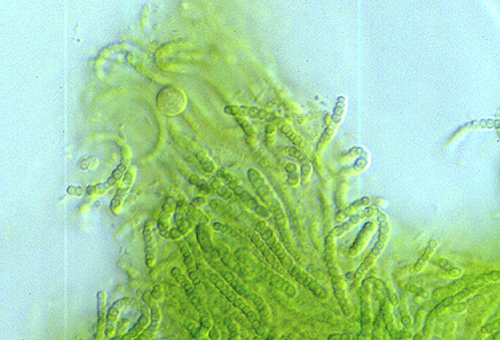
Mat Community Knit strings of cyanobacteria from green layer of a microbial mat from Great Sippewissett Saltmarsh, Falmouth, MA
Mat Community Cross-section of a microbial mat from Great Sippewissett Saltmarsh, Falmouth, MA
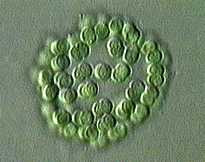
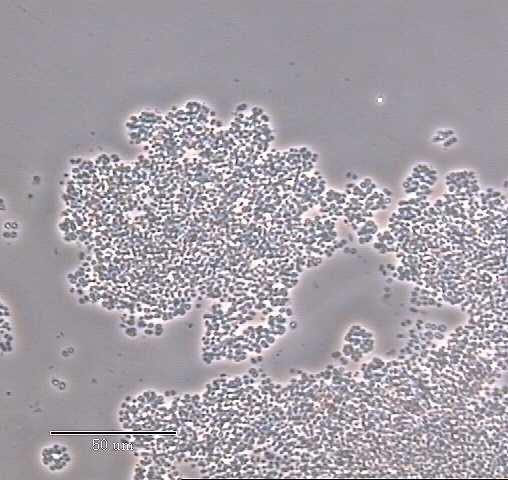
Merismopedia - Colony of Merismopedia (Agmenellum [Drouet], Synechocystis Low GC cluster [Waterbury and Rippka]). 200X

Merismopedia elegans, 640X


Microcoleus sp. - One of those in the anomalous Microcoleus/Symploca/Lyngbya/Take-a-guess range. 400X
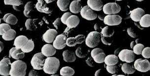
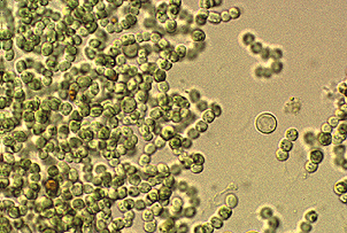
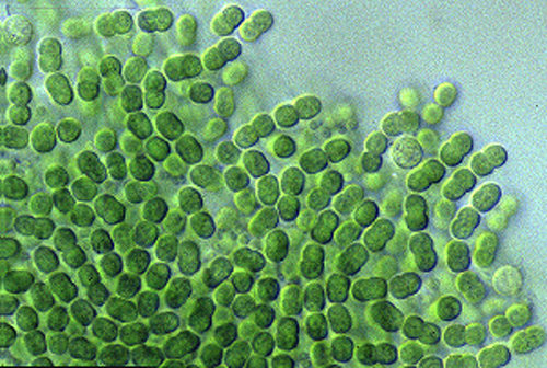
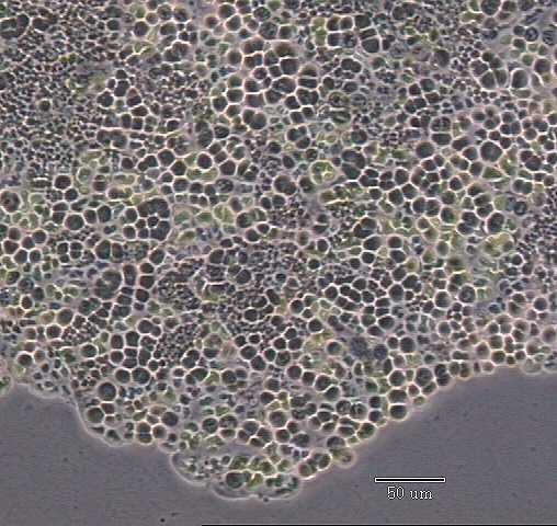
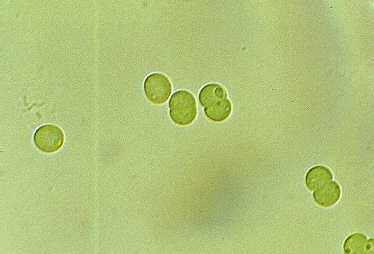
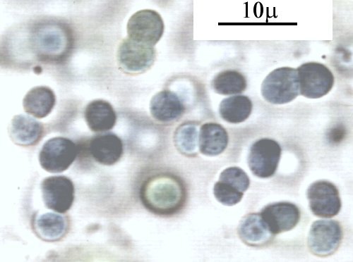
Microcystis aeruginosa enlarged 2500X
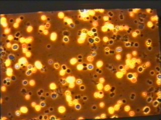
Microcystis aeruginosa strain PCC 7806. Autofluorescence image.

Microcystis aeruginosa strain PCC 7806. Autofluorescence image.
Microcystis aeruginosa strain PCC 7806. Autofluorescence image.
Microcystis aeruginosa strain PCC 7806. Autofluorescence image.
Microcystis aeruginosa strain PCC 7806. Autofluorescence image.
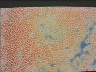
Microcystis aerogenosa strain 1450/10 in very old (6 month) stationary phase cultures imaged with autofluoresence. 1000X
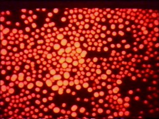
Microcystis aerogenosa strain 7806 in very old stationary phase cultures imaged with autofluoresence. Note transient "bright strike" feature. 1000X
Microcystis aerogenosa strain 7806 in very old (100 d) stationary phase cultures imaged with phase contrast. Note multi-planar division. 1000X

Microcystis aerogenosa strain 7806 in very old (100 d) stationary phase cultures imaged with phase contrast. Note narrow sheath. 1000X
Microcystis aerogenosa. 100X
Microcystis sp. bloom
Microcystis sp. - Becoming confluent. 200X
Microcystis sp. bloom from bird's eye view
Microcystis aeruginosa bloom, Lake Mokoan, Victoria, Australia.
Microcystis sp. - Separate colonies. 200X
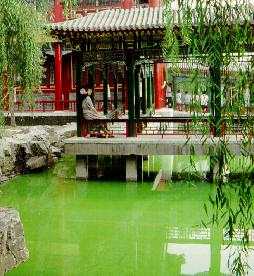
Microcystis sp., bloom, Grandview Garden Park, Beijing
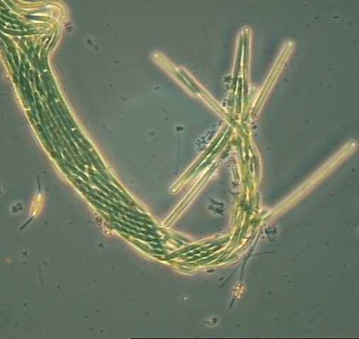
Mixture - Oscillatoria cf. chalybea, Planktothrix agardhii, and Anabaena spiroides. 200X
Mixture - Oscillatoria cf. chalybea, Planktothrix agardhii, and Arthrospira jenneri. 200X
Mixture, including Thiothrix and Oscillatoria sp.
Mixture, including Spirulina and Microcoleus sp. and diatoms together with Thiopediasp.
Mixture of cyanobacteria from microbial mat community
Mystery Bug - Truly strange. Spirulina major inside a bundle. 400X

Mystery Bug - Unidentified filament. Is it made of diatoms, desmids, cyanobacteria, or the unknown? 400X

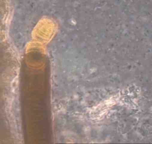
Mystery Bug A - Planktothrix agardhii with a swollen terminal cell. 200X. For Mystery Bugs A through E, it has been suggested that the swollen terminal cells are fungal (chytrid) fruiting bodies (sporangia) as hyphae have been observed running the length of similar trichomes.
Mystery Bug B - Planktothrix agardhii with a double swollen terminal cell. 200X. See Mystery Bug A.

Mystery Bug C - Another odd Planktothrix agardhii. 400X phase contrast. See Mystery Bug A.
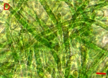
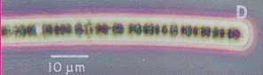
Mystery Bug D - Another odd Planktothrix agardhii. 400X phase contrast. See Mystery Bug A.

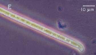
Mystery Bug E - Same as above without phase contrast. 400X. See Mystery Bug A.
Mystery wavy cyanobacteria
Mystery cyanobacteria in a colony

Mystery cyanobacteria showing vessicles


Nodularia spumigena enlarged 1250X
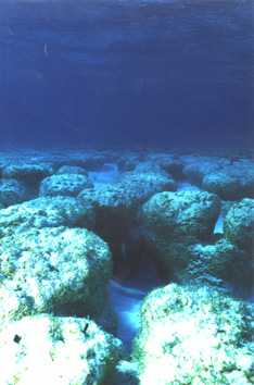
Nodularia sp. bloom in situ from an undersea porthole in the Baltic Proper
Nostoc (Anabaena) azollae - Heterocyst clearly visible. 400X
Nostoc (Anabaena) azollae - Numerous akinetes visible. 400X

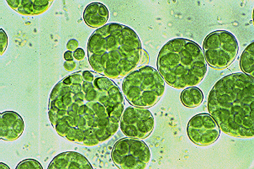
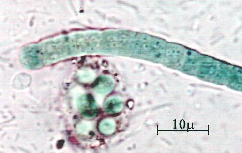
Nostoc sp. - Possible germling. 400X
Nostoc sp. - Possible colony or is it a strange type of Anabaena spherica?. The only thing for sure is that nothing is for sure. 200X
Nostoc sp., 130X
Nostoc sp. at 10X

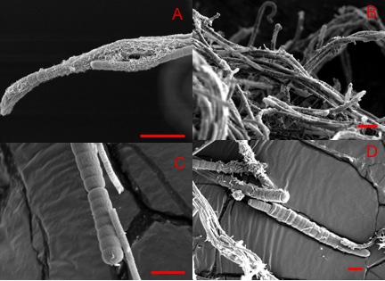
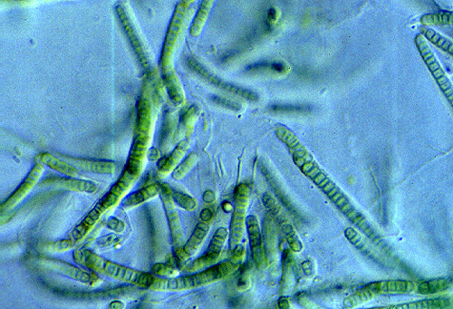
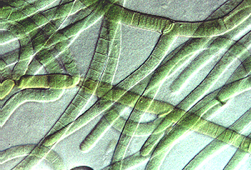
Nostochopsis sp., cell and branching
Nostochopsis sp., gross structure
Nostochopsis sp., detailed image
Nostochopsis sp., pediellate heterocyst
Nostochopsis sp., intercalary heterocyst
Oscillatoria agardhii - Autofluorescence image.

Oscillatoria cf. chalybea - With Planktothrix agardhii, and possible Raphidiopsis sp. 200X
Oscillatoria "chlorina" - Quotes represent the contributor's resentment at identifying cyanobacteria using color. 400X
Oscillatoria geminata - 400X
Oscillatoria limosa, 640X


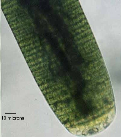
Oscillatoria princeps - 200X
Oscillatoria princeps - 100X
Oscillatoria princeps - Social gathering with some friends. 200X
Oscillatoria splendida - 400X phase contrast

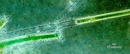
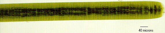
Oscillatoria sp. - The famous "big Oscillatoria". Species not known. It may be undescribed. 200X
Oscillatoria sp. - Note the granules in the terminal cell, along with its thickened membrane. 400X
Oscillatoria sp. - With the bright light rendering its striking granules more visible. Note the spiral arrangement of the granules. 100X

Oscillatoria sp. - The same specimen as above. 200X

Oscillatoria sp. - At low magnification. 40X
Oscillatoria sp. together with a Synechocystis microcolony. Note the numerous bacteria in the background.
Oscillatoria sp., 640X
Oscillatoria sp., likely a hormogonium, from a pulp and paper waste-treatment system in Brazil


Phormidium retzii - Tentative identification. 200X
Phormidium uncinatum - Tentative identification. 400X phase contrast
Phormidium uncinatum - Same as above without phase contrast. 200X
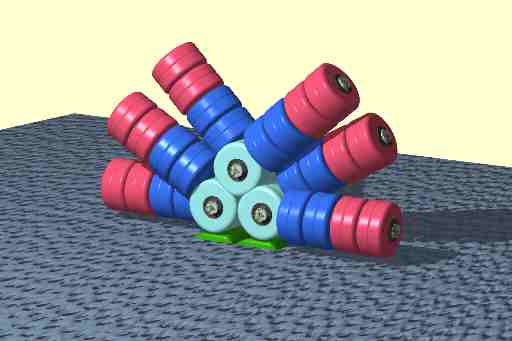
Phycobilisome diagram Each disk in the phycobilisome structure represents a hexameric aggregate, (alpha-beta)66, of a phycobiliprotein complexed with a linker protein: Phycoerythrin = red, Phycocyanin = blue, Allophycocyanin = light blue. The green elements are the two PSII reaction centers that associate with each phycobilisome.
Planktothrix (Oscillatoria) agardhii - With visible pseudovacuoles. 400X phase contrast

Plankothrix agardhii - microcystin-containing bloom in a Scottish freshwater lake
Plankothrix agardhii - shoreline deposit of a microcystin-containing bloom in a Scottish freshwater lake
Planktothrix rubescens. 100X.


Raphidiopsis curvata - Mixed with some straight chains that are either more Raphidiopsis or Cylndrospermopsis raciborskii without akinetes or heterocysts. 400X
Raphidiopsis curvata - Same as above with phase contrast
Raphidiopsis cf. mediterranea - Could be R. brookii. 400X

Raphidiopsis curvata - 200X
Raphidiopsis cf. mediterranea - Pseudovacuoles clearly visible. 400X phase contrast


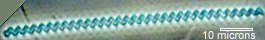
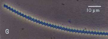
Spirulina major - 400X phase contrast


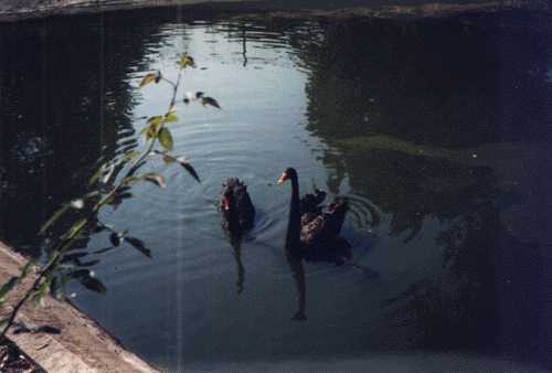
Spirulina sp. (Arthrospira) culture contaminated by brown swans in South Bend, IN.


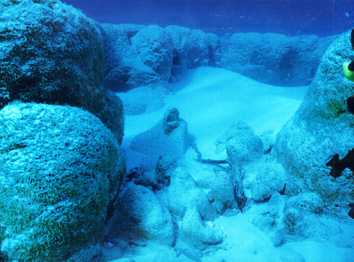
Columnar Stromatolites and Thrombolites from tidal channel at Lee Stocking Island, Bahamas
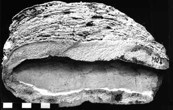
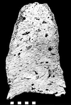
Stromatolite Cross-section, columnar procaryotic stromatolite grown on Calianassa burrow. Bar = 1 cm.
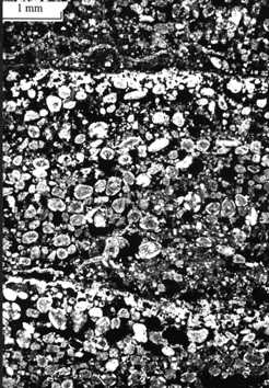
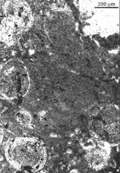
Stromatolite Photomicrograph, thin section of prokaryotic stromatolite showing alternating layers of dense micrite and coarse detritus.
Stromatolite Photomicrograph, thin section of eukaryotic stromatolite showing undulose laminations formed by a combination of eukaryotic algal and cyanobacterial activities.
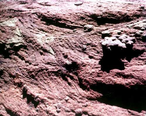
Stromatolites found in Middle Proterozoic formations of the Hakatai Shale in Grand Canyon National Park.
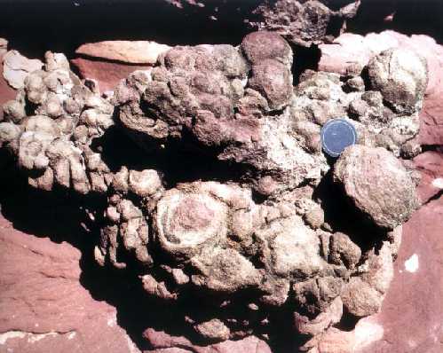
Stromatolites found in Middle Proterozoic formations of the Hakatai Shale in Grand Canyon National Park. Lens cap is 55 mm.
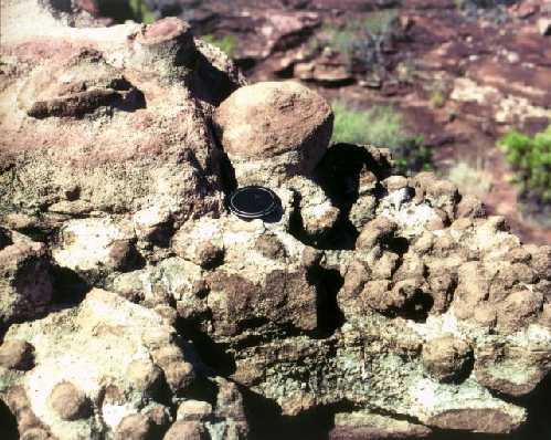
Stromatolites found in Middle Proterozoic formations of the Hakatai Shale in Grand Canyon National Park. Lens cap is 55 mm.
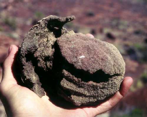
Stromatolites found in Middle Proterozoic formations of the Hakatai Shale in Grand Canyon National Park.

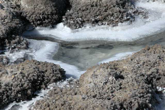
Frozen Stromatolites, Shark's Bay, Australia
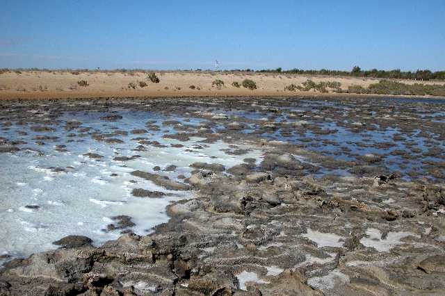
Frozen Stromatolites, Shark's Bay, Australia
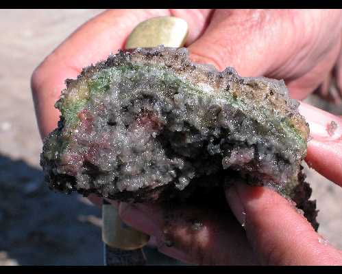
Frozen Stromatolite Mat, Shark's Bay, Australia
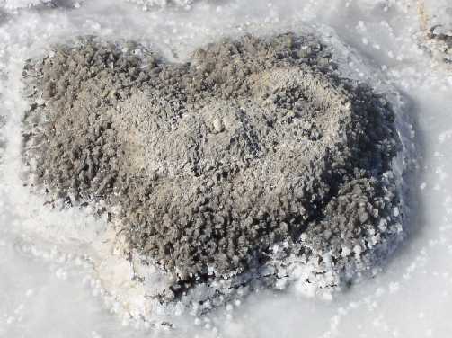
Frozen Stromatolite, Shark's Bay, Australia

Frozen Stromatolites, Shark's Bay, Australia
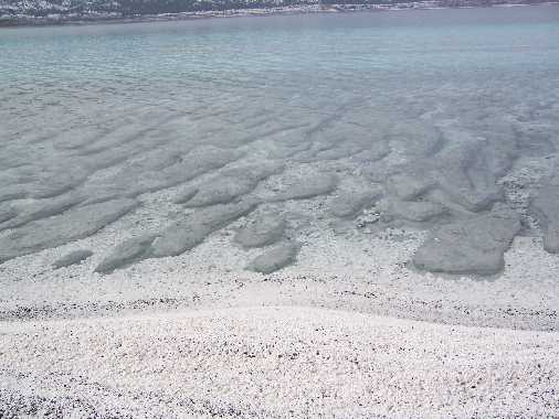
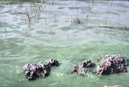
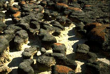
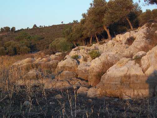
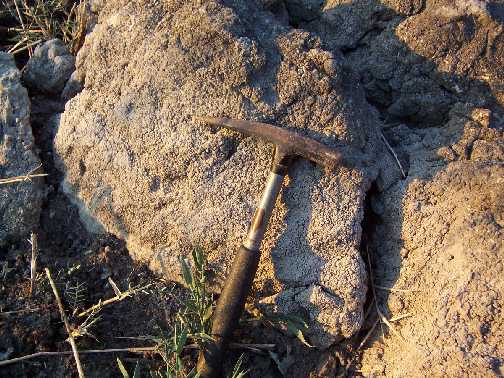
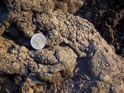
Living Stromatolites, Salda Lake, Turkey
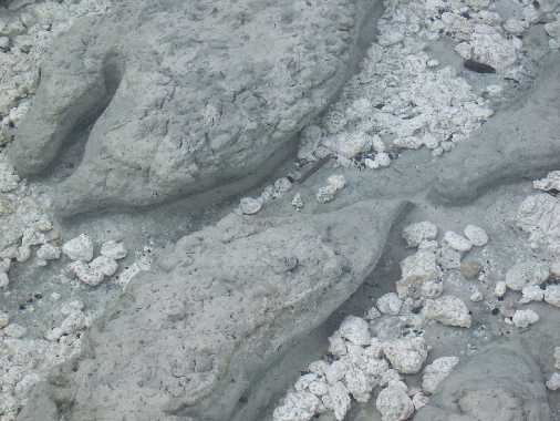
Living Stromatolites, Salda Lake, Turkey
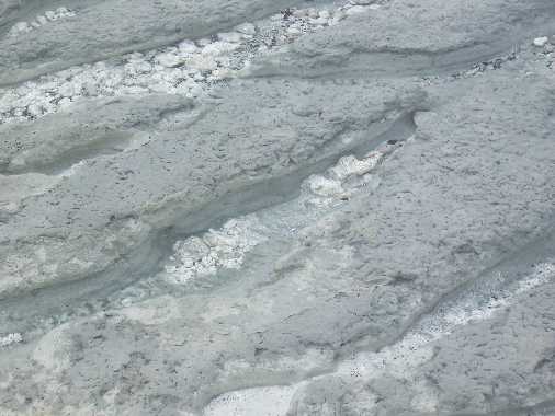
Living Stromatolites, Salda Lake, Turkey
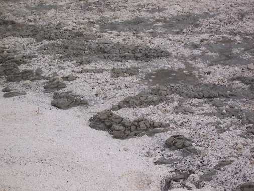
Living Stromatolites, Salda Lake, Turkey
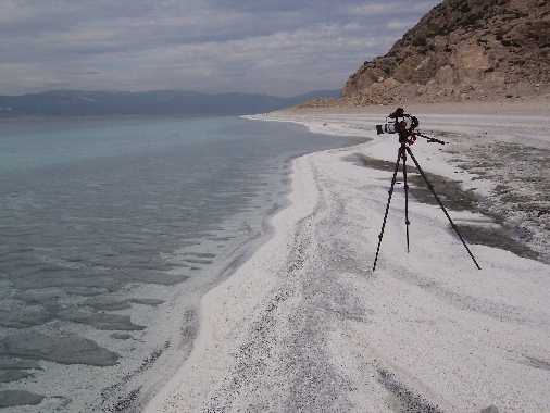
Living Stromatolites, Salda Lake, Turkey
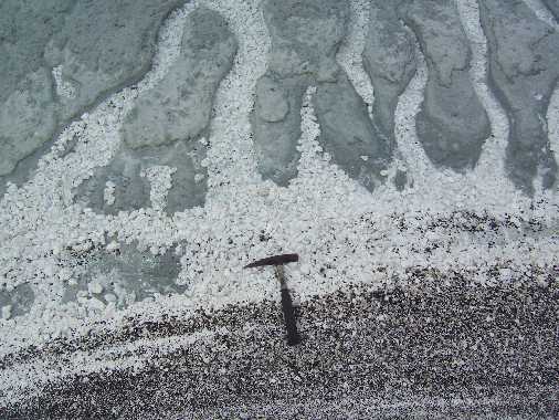
Living Stromatolites, Salda Lake, Turkey
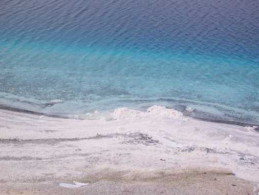
Living Stromatolites, Salda Lake, Turkey
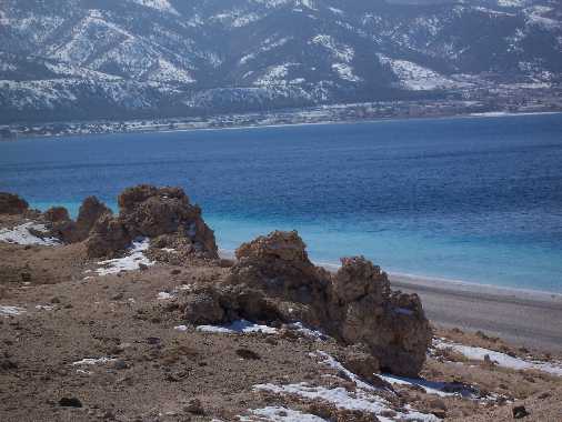
Living Stromatolites, Salda Lake, Turkey
Stromatoids, dry lake bed near Agaean Sea
Stromatoids, dry lake bed near Agaean Sea
Stromatoids, dry lake bed near Agaean Sea
Thrombolite Heads heavily overgrown by Lee Stocking Island, Bahamas
Thrombolite of modern origin from Lee Stocking Channel, Bahamas. Bar = 1 cm.
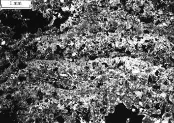
Thrombolite Photomicrograph, thin section showing boundary between micritic clot and detritus-rich sediment pocket
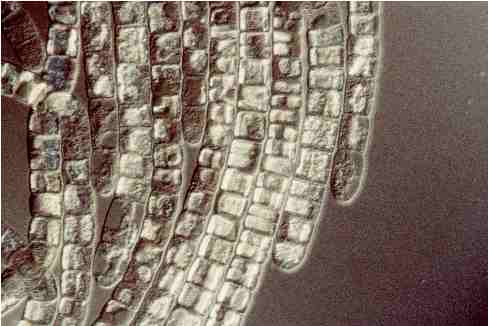
Trichodesmium sp., Nomarski optics, 1000X
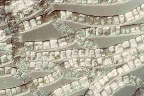
Trichodesmium sp., Nomarski optics, 1000X
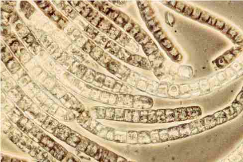
Trichodesmium sp., Phase contrast, 1000X
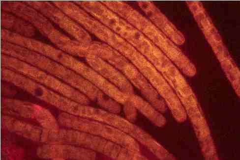
Trichodesmium sp., Fluorescence optics, 1000X
Trichodesmium sp., Fluorescence optics, 1000X
Trichodesmium sp., Phase contrast, 1000X
